HOME HYGIENE LIBRARY CATALOG TABLE OF CONTENTS GO TO NEXT CHAPTER
We are frequently asked, "Why do you presume to say what is natural to the system--how do you judge what is poisonous?"
Nature answers these much disputed questions, as she does so many others, in the iris of the eye. Substances congenial to the body, those which in quality and quantity normally belong to it, do not show in the iris. But all substances foreign or poisonous to the organism may reveal their presence and location by certain well defined signs and discolorations in the corresponding areas or fields of the iris.
For hundreds of years physicians and scientists have disputed as to the advisability of introducing into the human body in foods or medicines, mineral and earthy elements in the inorganic mineral form. Homeopaths, eclectics and physio-medical physicians seceded from the allopathic school of medicine because they condemned the use of inorganic minerals in physiological doses as medicines.
Nature Cure advocates, from the very beginning of the movement, strongly emphasized the fact that even those minerals and earthy elements which naturally belong to the human body must be taken as food or medicine only in the live, organic form in vegetable or animal protoplasm.

They claimed that iron, lime, sodium, potassium, magnesium, sulphur and phosphorus, though they are of the greatest importance in the vital economy of the body, must not be taken in the inorganic, mineral form, lest they accumulate and concentrate in certain parts or organs of the body for which they exhibit a special affinity and there become harmful and dangerous to health and life. The student should carefully study Vol. I., Chap. XXIV., in which this subject is fully treated.
Previous to the discovery of Nature's records in the iris, the question as to whether inorganic minerals and earthy elements are beneficial or harmful to the human organism, was largely a matter of opinion and controversy. No positive proof in support of either position was available.
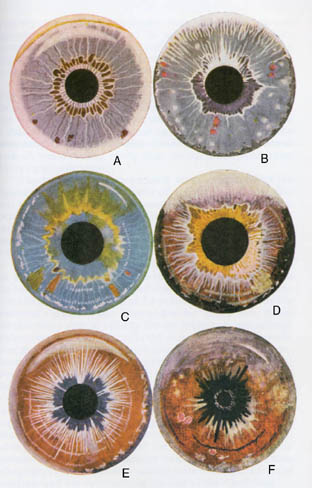
Iridology now proves beyond the shadow of a doubt that these substances, when taken in the inorganic form, accumulate in certain parts or organs for which they have a special affinity, as shown in color plate, page 116.

Before beginning the description of the various color signs in the iris I must call your attention to the fact that the iris of perfectly normal human beings exhibits only one of two normal colors--a light azure blue for all pure blooded descendants of the Keltic and Indo-Caucasian races, and a pure light brown for the first three Aryan subraces and for the descendants of the fourth or Atlantean root race. (The subject of racial characteristics has been briefly treated in Chapter IV.)
From this it follows that any other color pigments or discolorations of the iris indicate either abnormalities, disease processes or the presence of foreign matter or poisons in the system. The Iridologist, therefore, carefully studies every color pigment in the iris and endeavors to discover its significance. While in this way a great deal of positive knowledge has been acquired concerning disease processes and the presence and exact location of foreign and toxic materials in the system, much remains to be explained in this intensely interesting field of scientific research.
The effects upon the system of the various poisons exhibited in the iris are described elsewhere in this volume under the respective drug headings. The irides on this color plate represent right eyes only. It is interesting to note that the pigments in the iris closely resemble the natural color of the corresponding drugs.
Fig. a.--Blue eye. This is a typical drug eye. The dark blue underground is covered with a whitish film produced by coal tar poisons, such as salicylic acid and creosote, and other poisons. The crescent in the upper margin of the iris is the arcus senilis or gerontoxon. In medical works it is described as an opacity of the upper margin of the iris. It is usually observed in people of advanced age, therefore the name--arch of the aged. It is supposed to be a sign of lowered vitality and resistance of the organism as a whole and the brain tissues in particular. We frequently notice similar encroachments of the cornea on other sections of the iris. They are indicative of a weak lymphatic condition of the tissues and of low vitality. The arcus senilis must not be mistaken for certain drug signs which are described in this volume.
The inner margin of the arcus shows a yellowish discoloration caused by some drug poison, probably quinin. In the upper half of the iris, in the brain region, is displayed a broad white crescent, the sign of potassium bromate and of other bromin combinations. The color of the crescent varies somewhat according to the various chemicals associated with bromin in medical prescriptions. Sometimes the crescent covers the upper border of the iris and is extended more or less around the outer margin. It then shows similarly to the sodium ring in the same iris picture.
The broad whitish deposit all around the outer margin of the iris was caused by the absorption of large quantities of sodium bicarbonate (common baking soda). A similar deposit is formed by various substances containing large amounts of sodium, such as sodium salicylate, sodium sulphate, sodium bromate, etc. The salts of lime, magnesium and potassium show in similar manner. The location of the "salt ring" indicates that these mineral elements tend to concentrate and locate in the outer muscular structures of the body and in the skin, also in the brain tissue where they may have a serious effect upon memory and mental processes.
The white wheel in the stomach region, directly around the pupil, indicates the presence of strychnin in this organ. The sign is easily recognized by its perfect circular outline resembling a wheel and the uniform structure of its tiny spokes. The strychnin wheel appears imperfectly in Fig. f, where it is overshadowed by the sign of atrophy of the digestive organs.
The area of the intestinal tract surrounding the strychnin sign shows the rust brown discoloration of iron. In many instances this characteristic iron rust pigment covers the entire gastro-intestinal area. Inorganic iron has an astringent, benumbing effect upon the tissues. Its sign therefore indicates a sluggish, atrophic condition of the digestive organs. We frequently observe the sign in the eyes of people who have long used drinking water heavily charged with iron.
The sharply defined brown spots in the lower half of the iris are the itch or psora spots. They are the signs of suppressed scabies or of other itchy skin eruptions or eczemata as described in Chapter VIII. As time elapses after the suppression of psoric eruptions the itch spots grow darker in color until they become blackish brown.
Fig. b.--Blue eye. The bright red spots in this iris are the signs of iodin. We find them in a large percentage of human eyes (in civilized countries). They are of a brighter shade of red or brown than the psora spots, and their outlines are more diffuse.
The whitish "snowflakes", mostly visible in the lower half of this iris, are the signs of arsenical poisoning. Their presence in the spleen, as in this case, often signifies enlargement of the spleen and the serious symptoms that go with it. The white flakes of arsenic are easily distinguished from the lymphatic rosary in the outer margin of the iris, visible in the form of white flakes on the inner border of a portion of the scurf rim. The lymphatic or typhoid rosary is more plainly visible in Figures c and e.
The scurf rim is the dark deposit on the outer margin of the iris, it being covered in the upper part by the white deposit. Where it extends all around the iris, as in this case, it dates back to infancy and indicates conigenital scrofula or psora. If it appears only in parts of the iris, in crescent form, it has been acquired later in life.
The white deposit in the upper half of the iris, particularly the brain region, indicates the destructive effect of mercury and coal tar poisons upon the brain tissues, aggravated by bromids and the salts of other metals. We observe such bad demarcation and frazzled appearance of the upper rim of the iris frequently in the eyes of people threatened with or affected by apoplexy, insanity or paresis.
The purple discoloration in the area of the digestive organs indicates lead poisoning. We frequently find this in the eyes of printers, painters and others who work with lead. The white rays extending from the upper margin of the pupil to the brain region are the sign of opium, cocain or morphin poisoning. In the eyes of drug fiends the white rays extend from all around the iris, partly from the pupil, and partly from the sympathetic wreath.
The heavy, white sympathetic wreath surrounding the digestive area shows the effect of opiates and of powerful coal tar poisons, such as phenacetin and antipyrin, upon the sympathetic nervous system. In similar manner the sympathetic wreath may show the peculiar color pigments of other poisons, such as the yellow color of quinin, or the reddish color of iodin.
Fig. c.--Blue eye. This iris shows plainly the yellowish discoloration of quinin in the upper part of the iris, in the brain region, on the sympathetic wreath and in the region of the liver. The imposition of the yellow on the blue of the iris produces a greenish tint. This makes the "green" eye. While quinin has a special affinity for the brain and sympathetic system, we find its sign also in the areas of stomach, bowels, liver and spleen in the eyes of individuals who have taken considerable quantities of the drug for the treatment of malarial fevers, colds, hay fever and other forms of acute and chronic catarrh.
The outer rim of this iris shows the greenish ring of mercury. We found it difficult to reproduce this plainly. It requires some practice to distinguish it in the living eye. The small white crescent in the right upper margin of the iris indicates gummata--"softening of the brain"-- the result of the destructive action of the mercury on the brain tissues. Individuals who plainly exhibit this sign usually suffer from some form of paralysis and are approaching the end of their suffering.
The lymphatic or typhoid rosary shows plainly in parts of the outer margin of the iris. The fields of diaphragm and sexual organs exhibit the brownish signs of ergot, in this case administered for hemorrhage in childbirth. The triangle of the pancreas contains a psora spot. The person who exhibited this sign was in the last stages of diabetes.
An iodin spot shows in the lower back. It is partly surrounded by white, indicating that the poison is in process of elimination. The patient who exhibited the sign told us that during the eliminating crises he distinctly noted the iodin taste. Elimination took place largely through furuncles.
The lower part of the iris exhibits plainly several sections of white nerve rings.
Fig. d.--Brown eye. The greyish wash over the upper part of the iris indicates antikamnia poisoning. This is one of the coal tar products commonly used for the treatment of headaches, neuralgia, and neuritis of the head. Iridiagnosis plainly reveals the fact that the poison has a special affinity for the brain region, more so than other coal tar products. We have traced many cases of insanity to the effects of this drug on the brain.
The yellowish discoloration in the region of stomach and bowels may be caused by sulphur or by scrofulous elimination after the suppression of skin eruptions in childhood. It must not be confounded with the yellow color of quinin which appears only in spots and clouds.
The sympathetic wreath with its white rays shows somewhat similar to that of Fig. b; the explanation is the same.
The scurf rim is very broad and dark, indicating suppression of eczematous skin eruptions by metallic poisons.
The grey cloud in. the region of kidney, bladder and genital organs is caused by turpentine. This substance has a special affinity for the kidneys. The nerve rings in this iris are turning dark indicating that nervous irritation is becoming chronic.
The lymphatic rosary is visible in places through the heavy scurf rim.
Fig. e.--Brown eye. About two-thirds of the outer margin of the iris exhibits a heavy salt ring; the inner one-third is covered by an "acquired" scurf rim.
The nerve rings show white, indicating acute nervous irritation. The lymphatic rosary shows on the inner margin of the scurf rim. The large crescent in the brain region indicates deposits of bromids and other metallic salts.
The black discoloration in the digestive area stands for a sluggish, atrophic condition of the membranous linings of stomach and bowels, produced most probably by the use of opiates and powerful cathartics from early youth in the form of paregoric, calomel, etc.
Fig. f.--Brown eye. This is a typical mercurial eye. The bluish rim in a brown eye indicates the presence of the poison in the system and its destructive effect upon the cuticle. The patient received many inunctions of mercurial ointments. The destructive effect of the poison on the brain tissues is revealed by the white crescent near the right upper margin of the iris.
The upper portion of the iris is covered with the greyish wash of coal tar products. The paralyzing effect of mercury and other poisons on the digestive tract is revealed by the black color in the corresponding area. Only the inner margin of the stomach region shows faintly the strychnin wheel. Much strychnin was given to overcome the paralyzing effect of the mercury and other drug poisons.
The white rays emanating from the digestive area indicate opiates taken to deaden the pains of locomotor ataxia. The red spots edged with white are typical of potassium iodid. We frequently find them in the spinal area of mercurial patients. This patient being in the last stages of mercurial destruction, commonly called tertiary syphilis, the nerve rings show black, indicating the atrophic condition of the nervous system.
Greyish clouds in the region of the throat and in the lower margin show the presence of glycerin. The yellowish flakes in the lung region are caused by phosphorus which was administered in the form of nerve stimulants.
We find that iron, after it has been taken in considerable quantities in the inorganic form, shows in the areas of stomach and bowels as a rust brown discoloration which closely resembles the color of iron rust (Color plate, a). I have verified this sign in hundreds of cases in people who had absorbed iron in inorganic form in medicines or in water strongly impregnated with the mineral. Cases like the following have been of frequent occurrence:
Several years ago a lady came to one of our public clinics for diagnosis from the iris. The area around the pupil corresponding to the region of the stomach and intestines, showed a very heavy iron discoloration. I asked whether she had not taken the mineral in some form of drugs or patent medicines, but this she positively denied. Adroit quizzing finally brought out the fact that for several years she had used water from the iron spring in Lincoln Park. After forming this habit she had suffered much from constipation and indigestion.
I explained to her that these ailments were probably the result of the iron poisoning. Following my advice, she adopted a pure food natural diet and began a course of eliminative natural treatment. Within six months the iron sign had disappeared from her eyes and the digestive organs were in normal condition.
Allopathic Uses:
1. Externally on mucous membranes and broken skin as constringent and striptic against diffuse hemorrhages, catarrhal discharges and other inflammatory exudates.
2. Internally the non-astringent preparations are used as hematinics together with such drugs as influence the diseased conditions on which the anemia or debility depend, for instance:
3. Iron arsenate in chronic skin affections, particularly lupus, lepra, psoriasis, eczema, scrofula and syphilitic lesions.
4. Iron sulphate in chronic diarrhea, dysentery and passive hemorrhages accompanied by marked relaxation.
5. Iron bromid or iodid as tonic-alterative in atonic amenorrhea and chlorosis in young women.
6. Iron glycerophosphate and iron of manganese during convalescense, asthenic nervous conditions and rickets.
7. Iron valerianate, in hysterical complaints complicated with chlorosis.
8. Iron and quinin in malarial cachixia, cardiac disease and nephritis.
9. Antidote in acute arsenical poisoning, repeatedly administered in form of dialysed iron.
Accidental Poisoning:
1. Mineral waters.
2. Proprietary blood tonics.
Toxicology:
1. Unabsorbed and excreted as iron sulphid, coloring stools black.
2. Dyspepsia. Stubborn constipation.
3. Abdominal pain relieved by pressure.
Sodium shows in the eyes in the form of a white wreath in the outer margin of the iris. (See Color plate, p. 116, Figs, a and e)
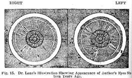
Before I became acquainted with Nature Cure, my eyes were heavily marked with drug signs. Some of these have entirely disappeared. Others are still faintly visible. Fig. 15 is a reproduction of charts of my eyes drawn fourteen years ago by Dr. Henry Lane, author of "Iridology".

It will be noticed that there is in the outer rim of the iris a broad white ring and a narrow inner ring. These signs were produced by inorganic sodium which for several years I had taken in large quantities to neutralize a hyperacid condition of the stomach.
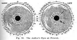
Today, as a result of natural living and treatment, these sodium rings as well as many other drug signs and disease signs have almost entirely disappeared, as shown in Fig. 16. The crosses in Fig. 15 indicate large iodin spots in liver and right kidney as they appeared sixteen years ago.
When I was a child our family physician had coated my neck at different times with iodin for the absorption of enlarged lymphatic glands. The poison had been absorbed through the skin and had accumulated in liver and kidney. This, together with decidedly unnatural habits of living, produced chronic ailments that incidentally led me into the work I am now doing.

Natural living and treatment have eliminated most of the sodium, as indicated in Fig. 16, but today, after forty-five years, the iodin spots are still faintly visible in several places, namely in left kidney, left lung and bronchi, and in right kidney and gall bladder. This proves that sodium and iodin, though congenial to the human body in the organic form, cannot be taken with impunity in the inorganic mineral form.
The history of my own case, as illustrated in these charts of the iris, shows that sodium is much more easily and quickly eliminated from the system than iodin. In this way iridology answers the question, "How long does it take to eliminate foreign matter and poisons from the system?''
We have observed that the symptoms of drug poisoning usually disappear much earlier than do the signs from the iris. This apparent contradiction can be accounted for by the fact that under the influence of natural living and treatment the general constitutional conditions are so improved that, although some of the poison is still present, the stronger organs are now able to compensate for the deficiency on the part of the weaker ones and therefore the effects of the drug poisoning are compensated for to some extent.* It may also be possible that drug poisons are more readily eliminated from vital organs than from the iris, on account of the more active metabolism in the former. (*See Chapter XXIV, entitled "Basic Diagnosis".)
As regards signs of those minerals and earthy substances which are naturally present in the human body, we find that iron, sodium, lime, sulphur and magnesium disappear much more quickly from the iris than iodin and phosphorus, indicating that the latter are more destructive and more difficult to dislodge.
Inorganic lime, magnesium and potassium show in the outer margin of the iris in the form of a grayish white wreath somewhat similar to sodium. (Color plate, p. 116, Figs, a and e.)
Only recently I examined a patient who came to us for diagnosis and treatment from far-away New Mexico. His iris exhibited very heavy sodium rings similar to my own as represented in Fig. 15, p. 122. Quizzing at first failed to reveal the source of the mineral accumulation in his system. Finally, however, it became apparent that the sign in the iris must have been produced by drinking for many years the water from the shallow wells of his native plains, strongly impregnated with alkali. These signs gradually disappear from the iris when the patient abstains from the use of mineral waters and adopts eliminative diet and treatment.
In the light of Nature's records in the iris, it is little less than criminal to give inorganic lime water, baking soda, iron, magnesium and table salt to little babes in artificial food mixtures when good cow's milk and fruit juices contain these minerals in great abundance in the live, organic (vitamine) form.
In the organic form in fruit juices and raw vegetable extracts, all these minerals may be taken continually in large quantities and will not show in the iris. An excess is easily eliminated from the system through the excretory organs, and we may safely say that the organism does not contain an excess of these positive mineral elements until a point is reached where the reaction of the urine is natural.
Thus Nature's records in the iris prove conclusively that she does not intend us to use these elements in the inorganic mineral form. The only apparent exception to this rule seems to be sodium chlorid, our common table salt. This might be explained by the fact that sodium chlorid is one of the ordinary products of kidney elimination, while other sodium combinations are not. There is no reason, however, why we should endanger health by using the table salt in such enormous quantities as is customary, since we can supply the demand of the system for sodium chlorid in the organic form by adding a liberal quantity of fruits and vegetables to our diet. There is no doubt whatever that table salt, when taken habitually and in considerable quantities, is very injurious to the system. The reasons for and against the use of salt have been fully discussed in the chapter entitled, "To Salt or Not to Salt", in the Nature Cure Cook Book, Vol. III of this series.
Sulphur, taken in the inorganic form, shows in the iris in the area of stomach and intestines in the yellow sulphur color. After absorption it has at first a stimulating effect upon these organs, but this is gradually followed by a sluggish, atrophic condition. Sometimes it is difficult to distinguish the sulphur color from the yellow color of quinin or the yellowish color of scrofulous elimination through the digestive organs.
HOME HYGIENE LIBRARY CATALOG TABLE OF CONTENTS GO TO NEXT CHAPTER