HOME HYGIENE LIBRARY CATALOG TABLE OF CONTENTS GO TO NEXT CHAPTER
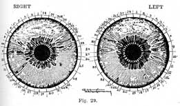
Mrs. V. was brought to us five years ago, in a dying condition. She had been troubled for twenty years with asthma, digestive disorders and many other ailments.
When she came to us the mucoid discharges from her throat were so copious and she was so emaciated that she presented the appearance of one laboring in the last stages of consumption.
For several months it seemed that the fatal crisis might come any day. The microscope showed some tubercle bacilli in the sputum, but not enough to make it a tubercular case.
After several months of natural treatment improvement came slowly but steadily. The healing crisis took the form of acute catarrhal elimination accompanied by low fever.
After seven months treatment she left for home in good condition. She felt fairly well for eight months; then overwork brought on another breakdown, and she returned to us for treatment.
The asthmatic attacks were very distressing, and she suffered greatly from an atrophic condition of the intestines--indigestion and malnutrition. For three months she could take very little food--not more than a few spoonfuls of milk or soft boiled egg with juicy fruit and fruit juices a day.
When conditions in the alimentary tract had greatly improved a serious crisis came in the form of an acute attack of pneumonia and pleurisy. In her already weakened condition this developed into a battle royal for life, but, as in all true healing crises, the healing forces came out victorious and from that time on she improved rapidly. After this last inflammatory crisis in the respiratory organs the asthma disappeared entirely.
Mrs. V. told us that her troubles had started in childhood with stubborn constipation, indigestion and malnutrition. For this she had received allopathic treatment. She remembered that she was given considerable calomel for the liver and bowels, and strychnin and arsenic as tonics to aid digestion.

When I first examined the patient her eyes distinctly showed the strychnin wheel in the stomach and the arsenic flakes in the outer iris, especially in the lungs.
These poisons, together with autointoxication and malnutrition due to her digestive troubles, probably brought on the asthmatic condition which followed in the wake of the medical treatment.
At the beginning of the asthmatic symptoms Nature tried to relieve the respiratory organs from the morbid encumbrances by a vigorous attack of pneumonia and pleurisy. This condition also was treated in the regular way with drugs and ice packs. From that time on the asthmatic attacks increased in frequency and severity.
In spite of, or probably as a result of, the continued medical treatment by the best specialists in Canada and the United States, her condition grew worse from year to year until life became a continual torture.
The sequence of healing crises, as well as her history and the records in the iris, revealed the causal chain in her case. While undergoing regeneration under the natural treatment she had to retrace the old acute diseases--the ailments that had been maltreated and suppressed in the past. The chronic conditions in the digestive organs, lungs and pleura had to become acute and run their natural courses before they could be permanently eradicated. For the last few years she has been practically free from the old complaints.
When I first examined her the areas of stomach and bowels were dark brown with many black spokes indicating an atrophic condition of the membranes and considerable destruction. The stomach revealed the strychnin wheel, while the intestines showed several iodin spots. She had been painted with iodin during the attack of pleurisy.
The bronchi, lungs and pleura showed chronic signs of the third and fourth stages. The brain region displayed the grey, mercurial crescent; the outer margin of the iris, the whitish flakes of arsenic. The entire iris was overspread with the greyish film of coal tar products. The scurf rim was heavy and continuous all around the iris; the lymphatic rosary also was very heavy, indicating the engorged and inactive condition of the lymphatic glands.
The iris pictures on this page show the appearance of her eyes when she first came to us five years ago.
The right iris shows a lesion in the region of the knee. In her girlhood the knee was injured by a fall on the ice. The right liver area shows the sign of subacute inflammation. The chronic signs in anus and rectum, left eye, stand for external and internal hemorrhoids.
At the time of writing this most of the signs just described have disappeared and the iris presents a clear, blue appearance. Of the drug signs only traces of mercury and iodin are visible.

When I first met Mr. B. three years ago he had a growth on the left side of his throat the size of a large walnut. It had a soft, red spot in the center which seemed ready to open. Several surgeons had diagnosed the case as true cancer and recommended immediate surgical removal.
The eyes of this patient at the time of my first examination, though apparently brown, showed on close examination a blue background. The brown, heaviest in the region of stomach and intestines, was superimposed.
When I mentioned this, he answered his mother had told him that in infancy his eyes were blue, but they had darkened and become brown when he was a few years old.
The scurf rim was heavy and dark except in the brain region. The darkening of the eyes and the formation of the scurf rim must have been caused through the suppressive treatment of skin eruptions, but this he did not remember and, his mother being dead, it was impossible to secure information on this point.
At the age of seven he suffered with inflammatory rheumatism. This was treated by an allopathic physician. He remembered that he was confined to bed for several months and that he did not fully recover from the attack for six months.
Two years later he was again prostrated with the same trouble and this time also he was not able to attend school for over six months. Since then he had been troubled periodically with rheumatism.
The treatment always consisted mainly in the administration of salicylates. This accounted for the heavy white ring in the outer margin of the iris, which stands for salts of sodium, magnesium, potassium and bromin, the bromin being more confined to the brain region.
We always find that people who have taken salicylates repeatedly and in considerable quantities exhibit in the digestive area of the iris the brown and blackish discolorations indicating atrophy of the membranes of the gastrointestinal tract. This patient was no exception to the rule.
On being questioned he admitted that since the first attack of rheumatism he had suffered from constipation and indigestion. These conditions had grown worse after the second attack and had become more chronic with advancing years. He reported that for many years he had never had a movement of the bowels without resorting to laxatives or enemas.
At the age of eleven he "caught the seven year itch", as he called it. This received the regular sulphur and molasses and blue ointment treatment. It proved a stubborn case and persisted in spite of drastic treatment for about six weeks.
Suppression of the scabies showed in the iris by several large itch spots, one in the right groin and one in the region of left neck, and another in right lower back. Several smaller itch spots showed in the intestinal tract.
During his childhood he was vaccinated a few times and received several antitoxin injections for immunization. This addition of disease matter to his system undoubtedly added to the vitiated condition of his vital fluids and helped to darken and discolor the iris.
From childhood up he was troubled, as before stated, with stubborn constipation, indigestion and malnutrition due to the atonic condition of the intestinal membranes. Catarrhal elimination through the membranous linings of the nasal passages, throat and bronchi endeavored to relieve the morbid condition of his system, but he did his best to prevent this by the use of cold and catarrh cures.
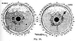
After his thirtieth year the rheumatism gradually became more chronic. Pathogenic obstruction in the system, together with the effects of the salicylates on the heart weakened that organ and caused it to dilate, which resulted in leakage of the mitral valve (Fig. 28, p. 239).
At the age of fortyone a swelling appeared on the left side of the neck. It was treated first with iodin; then several doctors pronounced it incipient cancer and recommended immediate surgical treatment. The patient balked at this for some time. When the further development of the growth left no doubt about its being of a malignant nature, he came to me for consultation and examination.
The first look in the iris revealed the large itch spot in the region of the left neck (Fig. 28). I explained to him what it meant--that the psoric taint together with general autointoxication of his system was undoubtedly responsible for the tumor. After a complete tracing of his ailments by the records in the iris from infancy on, he at once grasped the reasonableness of my explanation and submitted to thorough natural treatment.
A description of the many crises he passed through and their significance would fill a good sized volume. Suffice it to say that within two months after the commencement of treatment his bowels acted freely, and the skin and kidneys had become more alive and active.
The first crisis came in the form of acute catarrhal elimination, which lasted four weeks. The thirteenth week, the second crisis period, brought a severe attack of acute rheumatism. This lasted for about three weeks and was followed in the fourth month by fiery, itchy eruptions all over the body. Several eczematous patches appeared on the abdomen and discharged an acrid, watery fluid. The patient one day exhibited these ugly looking sores to a visiting physician who was interested in our work. The doctor could not understand why the patient seemed to be so much elated over his affliction until I explained to him that I had predicted the appearance of itchy eruptions as a form of healing crisis.
I also explained the significance of the itch spots; that they stood for suppressed psora and that this constitutional taint would have to work out through acute elimination before a reduction of the malignant growth could be expected.
It is now three years since the patient ceased taking treatment. The itchy eruptions appeared and disappeared periodically, extending over a period of six months. In the meantime the tumor in the neck softened and diminished in size slowly but steadily. As the vital fluids became pure and normal the food was taken away from the parasitic growth and pure blood and lymph gradually absorbed its pathogenic materials.
During the crisis periods the patient underwent three fasts of seven days, two weeks, and four weeks respectively. These, together with strict raw food and at times dry food diet, aided greatly in purifying the system of its pathogenic encumbrances.
Fig. 28 shows the records in his eyes as they appeared when I first examined him. Note the heavy scurf rim, partly covered by the salt ring, the dark brown discoloration and black spokes in the gastro-intestinal area, standing for the atonic condition of the membranous linings of these organs caused by salicylates. The liver also shows dark, indicating a sluggish condition. The itch spots in groin, neck and intestines are plainly visible. They were dark brown in color, indicating that the suppression had taken place many years previously. The broad white ring in the outer iris stands for deposits of salicylates. A heart lesion is plainly visible close to the sympathetic wreath in the left eye. (Area 10.)
The upper part of the iris in the brain region shows the greyish veil of coal tar products. Iodin is visible in left throat. The left leg had been crushed in a railway accident, which is indicated by a diagonal closed lesion.
The causes and rational treatment of diabetes mellitus will be described in Vol. V of this series. In the following I shall confine myself to a description of the signs of the disease in the iris.
From the viewpoint of Natural Therapeutics we distinguish two forms of diabetes--the functional and the organic. The functional form of the disease is caused by pathogenic (mucoid) obstruction in the tissues of the body. Pathogenic obstruction prevents absorption of sugar by the cells in the muscular tissues and its combustion incidental to the performance of muscular labor.
Under consumption causes excessive accumulation of sugar in the circulation, and excretion through the kidneys. If this continues for a considerable length of time, it results in the degeneration of these organs through overwork and irritation by the sugar and poisonous by-products of glycosuria such as indican, acetone, diacetic acid, ptomains, leukomains and other pathogenic substances. From this we see that affections of the kidneys in diabetes are, as a rule, of a secondary nature, not primary. It explains why the most serious chronic lesions appear in the pancreas, liver, stomach and intestines, while the kidneys in the initial stages of the disease exhibit signs of acute irritation.
When the tendency to sugar excretion is due to pathogenic (mucoid) obstruction in the tissues of the body, then the lower half of the iris usually appears darkened while the upper half shows whitish. This indicates that the circulation is impeded in the surface, extremities and muscular tissues of the body, while congestion exists in the larger internal arterial blood vessels in the brain, lungs and heart, giving rise to high blood pressure. In the advanced stages of the disease this is followed by weakness of the heart muscles or atony of the cardiac and vasomotor centers resulting in low blood pressure. The intestinal area is usually very much distended and shows dark discolorations.
The organic form of the disease is due in most cases to disease of the pancreas. The liver is the sugar refinery and sugar storage house of the body. During periods of excessive production and under consumption it stores sugar in the form of glycogen and releases it when needed as fuel material for the production of heat and muscular energy. The sugar liberating activity of the liver is regulated and retarded by certain as yet obscure secretions of the pancreas; in other words, the pancreas in this respect acts as a brake on the liver. If the brake or regulator is out of order the liver issues more sugar than needed. The excess accumulates in the circulation and gives rise to glycosuria or diabetes mellitus.
Abnormal conditions of the pancreas are plainly visible in the iris in a triangular projection from duodenum and cecum. If the organ is normal there is nothing to be seen in the corresponding region of the iris. If it is abnormal we notice a triangular bulge of the intestinal wreath projecting into areas 13 and 14, right eye. The typical appearance of this sign is illustrated in Figs. 13-18-22-24.
In this triangle we find portrayed the various signs of pancreatic diseases. In many cases I have observed the signs of acute or chronic inflammation; in others, the signs of suppressed itch. (Color plate, page 116, fig. c.) In some instances drug poisoning or suppression of psoric skin diseases dated back to early infancy. Frequently such patients strenuously deny having had itchy eruptions or eczemata or having taken the drug shown in the iris, but careful inquiry from relatives or the family physician elicits the fact that the drug had been administered for some infantile ailment, or the skin eruptions had been suppressed during the first years of life. It takes but very little poison to affect the tender organism of an infant. In many instances a few doses may be sufficient to affect an individual for life.
The diagnosis from the iris is especially valuable for detecting diseases of the pancreas. Though frequently diseased, it is hardly ever mentioned in allopathic and osteopathic diagnoses. The pancreas is overlapped by the stomach and intestines, therefore if it gives any subjective symptoms of discomfort or pain, these are usually attributed to affections of the stomach or of the intestines, while the signs in the pancreatic triangle in the iris reveal the true nature of the trouble.
Albuminuria as well as diabetes is primarily not a kidney disease. Both ailments may be caused by degenerative changes in the kidneys, the filter organs, resulting in leakage of sugar and albumen from the blood stream. But in the majority of cases the trouble is due to abnormal constitutional conditions. As explained under diabetes these may be functional or organic.
The initial stages of Bright's disease are usually caused by pathogenic obstruction of the capillary circulation and intercellular spaces. This interferes with the osmotic processes of nutrition and drainage. It prevents the consumption of proteid food materials and causes their accumulation in the blood stream, necessitating their discharge through the kidneys.
Pathogenic obstruction is gradually followed by degeneration and decomposition of the proteid constituents of cells and tissues and their absorption by the blood and lymph streams. The destruction of cellular protoplasm is undoubtedly hastened by systemic acids and by drug poisons, and as it proceeds the functional stages of the disease change into the organic or destructive stages involving also the kidneys.
Pathogenic obstruction is indicated in the iris by general darkening of the color, heavy scurf rim, white signs of acute inflammatory processes, darkening of the digestive area, nerve rings, etc. Organic destruction of tissues and organs caused by pathogenic obstruction and by the action of systemic and drug poisons is indicated by the signs of the third and fourth stages of disease.
Fig. 25, p. 226, shows chronic deterioration in both kidneys in a case of albuminuria in the advanced stages.
The female sex organs are much more complicated and therefore more prone to disease than the male organs. Most of the ordinary diseases of the female sex organs have been described in Chapter XVII, entitled "Woman's Suffering", in Vol. I of this series. In this chapter I shall confine myself to describing those diseases of the sex organs which are directly or indirectly due to venereal or gonorrheal infection.* (*This subject lias been treated more fully by the author in a booklet entitled "The Black Stork".)
The allopathic school teaches that these diseases are in themselves of a chronic, destructive nature, and that their progress must be stopped as soon as possible by local and constitutional treatment, by means of drugs, cauterizations, surgical operations, etc. These teachings and practices are erroneous and destructive. We have proved in many hundreds of cases that these diseases are, in themselves, of the acute inflammatory type, that when naturally treated they run a normal course through the five stages of inflammation as described in Vol. I, and then leave the system in a cleaner and more normal condition than it was before the infection.
Not a single one of these cases treated by us (before suppression had taken place) during the last seventeen years has exhibited secondary or tertiary symptoms. As I have explained many times, it is the suppression of these diseases during the acute and subacute stages by the allopathic treatment that creates the chronic stages and loads the system with destructive drug poisons which are responsible for the so called tertiary stages of syphilis and the worst kinds of other chronic destructive diseases.
Allopathy looks upon syphilis as more serious in its after effects than gonorrhea. Practical experience teaches us that the reverse is true. The average gonorrheal case exhibits much more painful symptoms and is more dangerous to the neighboring organs, as well as more destructive in its chronic after effects than syphilis. The only reason why syphilis is followed after a lapse of years by locomotor ataxia, paralysis agitans, paresis, and a multitude of other so called tertiary diseases is, that slow acting but powerful, insidious poisons are used for its suppression.
The gonorrheal acute catarrh of the membranous linings of the urethra and the syphilitic ulcer are slightly differing manifestations of the same venereal disease. This was acknowledged by Dr. Frankel, an allopathic specialist and writer on sexual diseases. He wrote: "The nature of the contagious poison is of minor importance. Everything depends on the more or less favorable soil the poison finds for development in the body."
It happens that a gonorrheal infection causes syphilitic symptoms or what is called a mixed infection of both gonorrheal and syphilitic symptoms and vice versa. Nature Cure physicians claim that persons with good skin action (light scurf rim) are more prone to the gonorrheal form of the disease, while those with low vitality, poor skin action and of psoric constitution tend to the syphilitic form of the disease. This I have been able to verify in many instances.
In "The Black Stork" I quote at length from the writings of Dr. Joseph Hermann, who has proved, not only theoretically but by thirty years of actual practice in one of the greatest hospitals for venereal diseases in the world, that neither gonorrhea nor syphilis are constitutional diseases; that they are easily curable in the acute stages by natural methods of living and treatment; and that all chronic and congenital after effects can be wholly avoided.
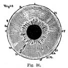
Fig. 30 illustrates a typical case of gonorrhea suppressed by injections of metallic poisons and internal medication. Area 20, urethra, and area 22, prostate gland, show the signs of subacute and chronic inflammation. As in many other cases of gonorrheal suppression the patient is now suffering from chronic prostatitis, and the urine has to be removed by catheters. His allopathic advisers insisted upon immediate operation. This would have meant greater suffering and the beginning of the end.

The suppression drove the disease taints and drug poisons into the bladder and kidneys; as a result the urine shows pus and albumen. Ever since the disease entered upon the subacute and chronic stages the patient has been impotent. This is indicated by the chronic sign in area 15, testes. During the subacute stages the right wrist became affected with gonorrheal arthritis; this also was suppressed and left the joint in an ankylosed condition (area 12). It is a peculiarity of gonorrheal arthritis that it affects only one joint at a time. The gonorrheal taint in the system will aggravate any tendency to rheumatism and make it more malignant. This type we call gonorrheal rheumatism.
Suppression of the acute catarrhal elimination from the urethra resulted in chronic catarrh of the nasal passages and the bronchi. This, in turn, was treated and suppressed for years with coal tar poisons. As a result of long continued drug poisoning, especially by salicylates administered for rheumatism and arthritis, the area of stomach and bowels shows chronic catarrhal signs, indicating indigestion, chronic constipation, gas formation and malnutrition. Of special interest in this case is the quinin sign in the brain region especially prominent in area 2, right cerebellum, which is the seat of sex life, the emotional nature, etc. The patient confided to me that from early youth he had suffered with excessive excitation of the sex impulse. Undoubtedly this was caused by quiuin taken in considerable quantities for chills and fever during his sixth and seventh years.

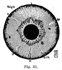
Fig. 31 illustrates the right eye of a woman fifty years old, who, at the age of twenty-five, contracted a syphilitic infection from her husband. The doctor who treated her, in order to shield the husband, did not inform her of the true nature of the disease. Not until she studied natural methods of healing and became a drugless healer herself did she find out the true nature of her ailments. When she came to me for treatment she exhibited a hole in the palate as large as a dime, which communicated with the nasal passages. She could not speak, nor could she take solid food because it entered the nasal passages. This lesion did not develop until many years after the syphilitic ulcer had been suppressed with mercury and potassium iodid. The inguinal glands and ovaries had been affected at the time by swellings (bubo) and inflammation. When I examined her, both areas (15, 18) in the iris showed chronic signs and a large iodin spot in the right bladder. The patient informed me that after the suppression of the original acute condition she had lost sexual sensation. The fields of the lower extremities reveal the signs of sub-acute inflammation. This was treated for years as sciatic rheumatism with salicylates and painkillers (narcotics and opiates). Subsequent developments showed that the supposed sciatic rheumatism marked the first stages of locomotor ataxia caused by the action of mercury and potassium iodid on the lower spinal cord. The upper part of the iris shows distinctly the greenish crescent of mercury. Iodin is visible in areas 28, 23, 10. Area 29, neck, shows signs of subacute inflammation due to engorgement and inflammation of the lymphatic glands.
The brain region exhibits acute signs in cerebrum and cerebellum. The accompanying symptoms are frequent headaches and dizziness. The patient under natural treatment experienced great improvement. The hole in the palate healed over perfectly within four months time. Her general condition improved sufficiently within six months that she was able to resume her work as drugless practitioner.
HOME HYGIENE LIBRARY CATALOG TABLE OF CONTENTS GO TO NEXT CHAPTER