

| Skip navigation | ||
 |
 |  |
|
|
||
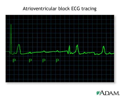
Atrioventricular block, EKG tracing

This picture shows an ECG (electrocardiogram, EKG) of a person with an abnormal rhythm (arrhythmia) called an atrioventricular (AV) block. P waves show that the top of the heart received electrical activity. Each P wave is usually followed by the tall (QRS) waves. QRS waves reflect the electrical activity that causes the heart to contract. When a P wave is present and not followed by a QRS wave (and heart contraction), there is an atrioventricular block, and a very slow pulse (bradycardia).
Update Date: 11/6/2006 Updated by: Glenn Gandelman, MD, MPH, Assistant Clinical Professor of Medicine, New York Medical College, Valhalla, NY. Review provided by VeriMed Healthcare Network.

| Home | Health Topics | Drugs & Supplements | Encyclopedia | Dictionary | News | Directories | Other Resources | |
| Copyright | Privacy | Accessibility | Quality Guidelines U.S. National Library of Medicine, 8600 Rockville Pike, Bethesda, MD 20894 National Institutes of Health | Department of Health & Human Services |
Page last updated: 02 January 2008 |