

| Skip navigation | ||
 |
 |  |
|
|
||
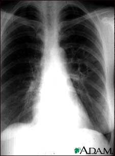
Coccidioidomycosis - chest X-ray

This chest x-ray shows the affects of a fungal infection, coccidioidomycosis. In the middle of the left lung (seen on the right side of the picture) there are multiple, thin-walled cavities (seen as light areas) with a diameter of 2 to 4 centimeters. To the side of these light areas are patchy light areas with irregular and poorly defined borders.
Other diseases that may explain these x-ray findings include lung abscesses, chronic pulmonary tuberculosis, chronic pulmonary histoplasmosis, and others.
Update Date: 8/10/2007 Updated by: Allen J. Blaivas, DO, Pulmonary, Critical Care, and Sleep Medicine, Department of Veteran Affairs, VA System, East Orange, NJ. Review provided by VeriMed Healthcare Network.

| Home | Health Topics | Drugs & Supplements | Encyclopedia | Dictionary | News | Directories | Other Resources | |
| Copyright | Privacy | Accessibility | Quality Guidelines U.S. National Library of Medicine, 8600 Rockville Pike, Bethesda, MD 20894 National Institutes of Health | Department of Health & Human Services |
Page last updated: 02 January 2008 |