

| Skip navigation | ||
 |
 |  |
|
|
||
Ultrasound comparison

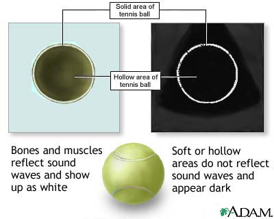
To demonstrate how an ultrasound works, imagine this tennis ball as an internal organ in the body. Like many organs, the tennis ball is solid on the outside and hollow on the inside. Solid structures, such as bones and muscles, reflect sound waves from the ultrasound transducer and show up as white in an ultrasound image. Soft or hollow areas, like chambers of the heart, do not reflect sound waves and appear as black. The white ring is the outer edge of the tennis ball being reflected back as an image while the center hollow area remains as black.
Update Date: 10/24/2006 Updated by: Stuart Bentley-Hibbert, M.D., Ph.D., Department of Radiology, Weill Cornell Medical Center, New York, NY. Review provided by VeriMed Healthcare Network.

| Home | Health Topics | Drugs & Supplements | Encyclopedia | Dictionary | News | Directories | Other Resources | |
| Copyright | Privacy | Accessibility | Quality Guidelines U.S. National Library of Medicine, 8600 Rockville Pike, Bethesda, MD 20894 National Institutes of Health | Department of Health & Human Services |
Page last updated: 02 January 2008 |