

| Skip navigation | ||
 |
 |  |
|
|
||
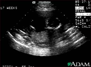
Ultrasound, normal fetus - ventricles of brain

This is a normal fetal ultrasound performed at 17 weeks gestation. The development of the brain and nervous system begins early in fetal development. During an ultrasound, the technician usually looks for the presence of brain ventricles. Ventricles are spaces in the brain that are filled with fluid. In this early ultrasound, the ventricles can be seen as light lines extending through the skull, seen in the upper right side of the image. The cross hair is pointing to the front of the skull, and directly to the right, the lines of the ventricles are visible.
Update Date: 7/26/2007 Updated by: Daniel Rauch, M.D., FAAP., Director, Pediatric Hospitalist Program, New York University School of Medicine, New York, NY. Review provided by VeriMed Healthcare Network.

| Home | Health Topics | Drugs & Supplements | Encyclopedia | Dictionary | News | Directories | Other Resources | |
| Copyright | Privacy | Accessibility | Quality Guidelines U.S. National Library of Medicine, 8600 Rockville Pike, Bethesda, MD 20894 National Institutes of Health | Department of Health & Human Services |
Page last updated: 02 January 2008 |