HOME HYGIENE LIBRARY CATALOG TABLE OF CONTENTS GO TO NEXT CHAPTER
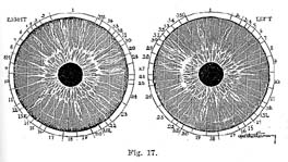
The area of the stomach is located directly around the pupil (A), that of the intestines surrounds the stomach (B) and the border of the intestinal field represents the sympathetic nervous system (C). See Chart, Frontispiece.

Acute conditions of these organs show white in the iris (fig. 17), while chronic conditions, accompanied by gradual atrophy of the membranes of these organs, create dark grey, brown and black discolorations. (Color plate, figs, e and f.)
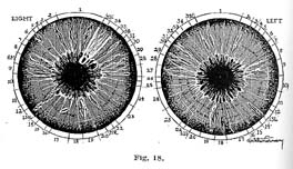
Acute catarrhal conditions of the stomach are usually accompanied by excessive secretion of hydrochloric acid as well as by systemic accumulations of uric and other acids (Fig. 17), while the chronic condition, accompanied by more or less atrophy of the membranous linings of the stomach, is responsible for deficient secretion of hydrochloric acid and pepsin (Fig. 18).
In order to find out whether the contents of the stomach are hyperacid or hypoacid, physicians of the regular school introduce rubber tubes into the stomach and take test samples of its contents for examination in the laboratory. The iridologist does not have to resort to this unpleasant and injurious practice. The showing in the iris reveals whether the subject is suffering from hyperacidity or hypoacidity. If the stomach area in the iris shows white, this is a sign of acute inflammatory and hyperacid condition, while dark discoloration indicates a sluggish, atonic or atrophic condition of the membranous linings and therefore a deficiency of hydrochloric acid and pepsin. As a result of deficient secretion the foods remain undigested, enter into morbid fermentation and create noxious gases and other pathogenic materials.
If the stomach, through long continued destructive processes, is in a condition of chronic inflammation or ulceration, the inner edge around the pupil shows dark and ragged. (Fig. 17, p. 215.)
As the disease processes in the stomach proceed from the acute and subacute to the chronic and destructive stages, we observe in the iris the appearance, first, of greyish or brownish spokes. These gradually darken until in the destructive stages they become quite heavy in appearance and black in color. (Fig. 18, p. 216.)
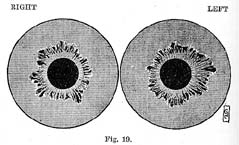
A weakened and relaxed condition of the digestive tract, resulting in enlargement and prolapsus of the organs is indicated in the iris by the distention of the areas of the stomach and bowels (Fig. 19, p. 218). This is true in a more pronounced degree of the intestinal tract, which is frequently greatly distended in the areas of the cecum, ascending colon, descending colon, sigmoid flexure and rectum, these parts of the intestinal tract being frequently enlarged on account of accumulation of food materials and feces.
What has been said about signs of acute and chronic conditions in the stomach is also true of the intestines. Frequently the stomach area appears whitish, greyish or light brown, while the intestinal area is enormously distended and shows the black spokes of chronic conditions. In such cases stomach digestion may be fairly active while the intestinal tract is in an atonic condition. (Fig. 19.)
In many cases of mercurial poisoning the intestinal area shows deep black, indicating the paralyzing effect of the poison on the intestinal membranes. The liver in such cases also shows chronic signs. The intestinal area frequently shows the signs of quinin, iron, sulphur, opium and its derivatives. We also find in the intestines the signs of iodin but not of strychnin. (Fig. 26, p. 227.)
Itch spots, the signs of suppression of psoric eruptions, we find quite often in the fields of the stomach and intestines. These always indicate a tendency to ulcers and benign and malignant tumors (Fig. 20). Cancer in these organs shows usually as a small black spot surrounded by white. (Fig. 14, Series IV, c.)
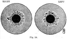
(Fig. 20, p. 219.) I examined this patient six years ago and found several large itch spots in the intestines. I informed the husband of the lady that these itch spots indicated a strong tendency to cancer, but she did not remain for treatment. Four years afterward he brought his wife to us in the last stages of cancer of the intestines. It was too late for recovery.
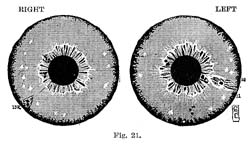
(Fig. 21, p. 220.) About the same time that I made this diagnosis I examined another lady whose left breast was slightly inflamed around the nipple. Her iris revealed itch spots in the left breast. I warned her also of the possibility of cancer. She took treatment for two months; then her husband, who did not believe in Natural Therapeutics, forced her to abandon the treatment and return to her home in a western city. When he brought her back three years later, the left breast was a solid mass of cancer. This case also had advanced beyond the possibility of improvement. Itch spots showed also in the left groin and liver. Mrs. S. remembered distinctly that in her childhood she had suffered with eczematous eruptions which were treated with "medicine and salves". Undoubtedly these remedies accounted for the heavy scurf rim in her eyes, and for the arsenical flakes.
If these cancer patients and their relatives had understood the nature of psora and its hidden clanger, both lives could have been saved.
I prefer to describe these organs together because they are companion organs and we find that when one of them is seriously diseased the other also is more or less affected.
These organs are the refineries of the body. The liver refines the end products of starchy and protein metabolism and discharges the waste materials thus extracted, partly in the form of bile, into the gall bladder and from there into the intestines, and partly in the form of urea which is excreted through the kidneys.
It has been known to medical science that, in addition to serving as a burial ground for dead red corpuscles, the spleen has much to do with the purification of the blood, but it was not clear just how this purification took place. Many theories have been advanced which have failed to withstand the tests of scientific research and clinical experience.
The new science of Natural Therapeutics for the first time gives a rational explanation of the true function of the spleen and of the lymph nodes in the lymphatic system. This new theory has been fully explained in Chapter IX, "Inflammation", Vol. 1, and also in connection with the study of various diseases.
According to the new version the spleen and the lymph nodes serve to filter the mucoid pathogenic materials out of the blood stream and to condense them into little compact bodies, the so called leukocytes or phagocytes which have been mistaken for live, germ killing cells.
The purpose of this condensation of pathogen, as elsewhere explained, is to render the blood serum more fluid and thus to facilitate its penetration into the intercellular spaces (osmosis) and thereby the nourishment of the cells by arterial blood and their drainage by way of the lymphatic and venous systems.
One of the principal reasons why Metchnikoff assumed that the leukocytes were germ killers was because they increased in numbers with the beginning of inflammation in any part of the system. He believed that they increased because more germ killers or phagocytes were needed to overcome the inflammation creating bacteria. The new science of healing proves that inflammation takes place on account of the increase in pathogen and leukocytes, which causes obstruction in the capillary circulation.
A number of serious diseases did not confirm the Metchnikoff theory. In miliary tuberculosis, malaria, typhoid fever, influenza and pernicious anemia the leukocytes are greatly reduced in numbers while the spleen and lymph nodes, the capillary circulation and the intercellular spaces are blocked with leukocytes and colloid (pathogenic) materials. (Fig. 12, p. 97, note lymphatic rosary.)
The following explains this phenomenon also: In these diseases the amount of pathogen in the circulation is so great that the trabeculae of the spleen and of the lymph nodes become so engorged with mucoid materials that they cannot any longer continue the pathogen condensing and filtering process. The failure of the spleen and lymph nodes to continue their normal functions explains the decrease of leukocytes in the circulation and the corresponding increase of colloid materials.
The enlarged spleen and swollen lymph glands in quick consumption, malaria, typhoid fever, influenza and pernicious anemia are in the same condition as a sieve that has become so clogged that it cannot any longer sift the solids from the fluids.
This crowding of the lymph nodes in the neck with colloid matter can be frequently observed after the extirpation of the tonsils and adenoids, which means suppression of colloid elimination. When the lymph nodes are thus engorged with pathogenic materials the surgeon knows no better than to cut them out. The practitioner of Natural Therapeutics promotes the elimination of the excess of pathogenic materials from the circulation and thus relieves the engorged spleen and lymph nodules.
The foregoing explains why in serious anemias and in the other diseases before mentioned we always find in the iris the signs of acute and subacute activity in the region of the spleen and usually also in the liver, because most of the diseases of the liver also originate in pathogenic engorgement. (Page 112, Series I.)
When the colloid or mucoid obstruction in these organs continues, the acute and subacute stages are followed by chronic and finally chronic destructive stages, which is readily explained by the fact that constant pathogenic obstruction interferes with nourishment and drainage of the cells and thereby brings about deterioration, degeneration and gradual destruction of the cells and tissues in the affected parts. (Page 112, Series III and IV, also fig. 22-13, right and left.)
Practically all diseases affecting a vital organ, whether it be the liver, spleen, kidneys, lungs, stomach or intestines, run the same course. Pathogenic obstruction first causes reaction by acute inflammatory processes. If these succeed in clearing the tissues of morbid materials, then follows recovery and normal function. (Page 112, Series I, B-E.)
If, however, the pathogenic encumbrances increase and become permanent, then the system is no longer able to remove the obstructions by acute inflammatory effort and the result is chronic degeneration and destruction. (Fig. 14, Series III, IV.) This again confirms the teachings of Natural Therapeutics, according to which acute disease is constructive or curative in tendency while the chronic stages are characterized by atrophy and destruction of tissues.
The color of the lesions in the iris, whether they be white or greyish, brown or black, indicates what stage of pathogenic development the disease has reached.
That the ancients understood the connection between diseases of the liver and spleen and emotional conditions is proved by the fact that the word "melancholia" means "black gall". Obstruction of the gall duct is frequently caused by the accumulation of colloid materials in the form of black, tarry accretions in the gall bladder. This interferes with the flow of the bile through the gall duct into the intestine, which in turn causes the surging back of the bile into the blood stream. The absence of bile in the intestines results in constipation. Bile in the blood stream irritates brain and nerve matter, causing mental depression, melancholia or hysteria. (Fig. 17 R, p. 215.)
I have been told on good authority that in over half the operations for gall stones no stones are found, but in place of them the black tarry substances before described--the "black gall" of the ancients. Such catarrhal obstruction of the gall duct may cause distention, painful symptoms and a bilious condition of the system, similar to that caused by obstruction by stones.
Engorgement of the spleen and of the lymph nodes results in excess of pathogenic materials in the circulation. These benumb brain and nerve matter, causing physical and mental lassitude, melancholia, insanity or, in acute diseases, mental depression, coma and death. (Fig. 18, p. 216.)
I examined Mr. L. shortly before he died of pernicious anemia. His eyes revealed an extraordinary combination of serious lesions and signs of drug poisons, as shown in Fig. 22. Lesions in the respective organs reveal a serious chronic destructive condition of the liver and gall bladder, accompanied by a collemic, engorged condition of the spleen. The brain region, as well as the areas of liver and spleen, show distinctly the color of quiuin. In his youth he had suffered for several years with malaria, which was treated with quinin in the orthodox way. Since that time he had been affected by melancholia, indigestion, chronic constipation, neurasthenia and many other troubles, making his life a continual torture until death relieved him at the age of forty-two. His eyes also showed subacute lesions in the kidneys, several segments of nerve rings, a chronic lesion in the right ear and a closed lesion in the left knee. The pancreas area shows a large psora spot.
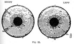
The brain region also revealed the presence of bromids and coal tar products which had been administered to suppress the headaches and insomnia caused by quinin poisoning.
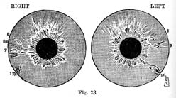
Fig. 23 shows a severe lesion in the spleen and a chronic catarrhal condition of the lungs. The patient had contracted "swamp fever" in South America. After he returned to the United States this was suppressed with quinin and arsenic. The pathogenic condition of the blood caused by the diseased spleen and liver was followed by incipient tuberculosis of the lungs.
Allopathic physicians admit that tuberculosis is much more dangerous when accompanied by spleen disease. Our theory of leukocytosis explains why this is so. "When the circulation is saturated with excessive amounts of pathogen this will engorge and obstruct the tissues of the lungs and thus prepare the soil for tubercular processes. This patient made a perfect recovery under natural living and treatment.
In practically all cases where patients told me that since they had the grippe they were troubled more or less with headaches, nervous irritability, insomnia, indigestion, bone aches, neuritis, physical and mental weakness, etc., I found in the iris the signs of quinin, indicating that the chronic after effects of influenza are not due to the disease itself but to its suppression by quinin and other poisons. This is also proved by the fact that in cases where influenza is treated by natural methods such chronic after effects never develop. Quinin shows plainly in the brain region.
The kidneys are especially prone to disease conditions because they are the filters of the system, whose function it is to eliminate from the blood stream all sorts of waste and morbid materials. As long as the system is in fairly good condition and the kidneys have nothing to cope with but the normal forms of waste matter such as urea and salts, they will remain in a healthy condition, but whenever they are forced to eliminate large amounts of pathogenic materials, earthy matter, uric acid and ptomains, bile salts, etc., the tender tissues of these organs become irritated and inflamed. This leads to morbid changes which in time render them incapable of performing their functions.
When acute and subacute irritation by poisonous excretions continues for too long a time, the tissues of the kidneys undergo degenerative changes. Microscopic examination of the urine then reveals kidney cells, tubules, casts, leukocytes, red blood corpuscles, pus cells and other debris of inflammatory breakdown.
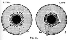
Fig. 24 shows both kidneys in a subacute condition. That this was not primary disease, is shown by the serious lesions in the pancreas and liver which were associated with diabetes in the advanced stages. There also showed in both eyes the signs of acute inflammation in the bladder. The stomach and bowel region revealed the dark signs of chronic catarrh, indicating an atrophic condition of the membranes of stomach and intestines, which resulted in malnutrition and systemic poisoning.
The history of the patient revealed that he had been suffering since early youth with malnutrition and constipation, which I traced to the use of paregoric and cathartics. The mother of the patient told me that she had been in the habit of giving her children those wonderful "soothing syrups" and "innocent laxatives" so as not to be disturbed at night or when engaged in her household duties. In the meantime the laudanum in the paregoric and the calomel was benumbing and paralyzing the liver and intestinal membranes of her children. At the age of twenty the patient developed diabetes. This was treated with the ordinary allopathic remedies, but instead of curing the disease the poison treatment resulted in chronic inflammation and finally in breakdown of the kidneys, accompanied by the discharge of albumen in addition to sugar. The patient died at the age of thirtythree, shortly after I had made the diagnosis from the iris of the eye.
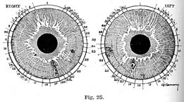
Fig. 25 shows chronic, destructive kidney lesions in both eyes. In the right kidney region as well as in the right back we notice large itch spots. There is a closed lesion in the left kidney and an iodin sign in the pancreas. The brain region shows the signs of acute inflammation.
The patient, when I examined her, was in the last stages of Bright's disease. The cause of the trouble is revealed by the itch spots. She remembered distinctly that she had had the itch several times in her youth and that it was suppressed with the usual remedies--sulphur and molasses and blue ointment (mercury). She had suffered from weakness of the kidneys and bladder ever since. The poison sign in the pancreas accounts for the presence of sugar.
The white lines all through the iris and the heavy nerve rings indicate irritation of the nervous system through uric acid and other pathogenic materials. In this case the concentration of the psoric taint in the kidneys as the result of the suppression of scabies ("seven year itch") was undoubtedly responsible for the gradual breakdown of these organs.
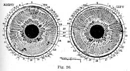
(Fig. 26.) These eyes indicate plainly a uric acid diathesis. The irritation caused by this systemic poison throughout the entire system is indicated by the white lines all over the iris, giving it a grey appearance. Irritation of the nervous system by uric acid and other systemic poisons is also indicated by the many prominent nerve rings. Uric acid in this case resulted in the forming of a stone in the left kidney. This was removed by a surgical operation four years before the patient came to us for treatment. Two years after the operation another stone had formed in the right kidney. Mr. S. told me that the new stone in the right kidney was giving him much more trouble than the previous one in the left kidney ; that for two years he had traveled from one hospital or sanitarium to another, his ailments growing worse all the time.
When one of a pair of organs is affected by constitutional disease and is removed by the surgeon's knife, the disease soon after manifests in the companion organ. This is of such common occurrence that anyone who runs may read, but the surgeons cannot or do not want to see that constitutional disease is never cured by the mutilation or extirpation of an affected organ. Common sense reasoning would tell us that the only way to meet the problem is to cure the constitutional disease back of the trouble--in the case under discussion, the uric acid diathesis. This can easily be accomplished by the natural methods of living and of treatment.
Before this patient came to us he developed at intervals of two weeks a violent inflammation of the affected kidney. This was accompanied by high fever and excruciating pains. The attacks, which were undoubtedly precipitated by irritation due to the stone in the kidney, would last about a week and then subside, to be followed after another interval of two weeks by another attack.
After he came under our care and treatment he had only one of these violent collicky attacks. After that he experienced only a few slight aggravations and improved rapidly. He left our institution five months after his arrival and, according to last reports, had not experienced another attack of kidney inflammation, but I learned from his friends that since that time he has been troubled a great deal with acute inflammatory rheumatism, which is to be accounted for by the fact that he was not at all strict in his adherence to the natural regimen of living. X-ray pictures which were taken just before his arrival and at the time of his departure showed that the stone had actually diminished one eighth of an inch in diameter in various directions.
The acute inflammatory attacks subsided so quickly because raw food diet and fasting reduced the hyperacidity of his system very rapidly, thus lessening the irritation caused by the stone and the pathogenic condition of the blood. The fact that the stone considerably diminished in size in five months' time proves that these calculi form only in blood highly charged with acid materials, and that they gradually dissolve under a normal alkaline condition of the vital fluids and eliminative treatment.
The digestive organs reveal a chronic catarrhal condition, also signs of iodin; in the outer margin of the iris, deposits of salts of sodium and magnesium; subacute catarrhal signs in bronchi, throat and nasal passages. The chronic lesion in the rectum stands for hemorrhoids.
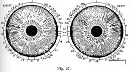
These are the eyes of a young lady who died from phthisis of the lungs at the age of twentyfour. I examined her eyes three months before her death, when it had become too late to save her life. The eyes showed the following lesions and other abnormalities.
One of the most prominent features is a very heavy dark scurf rim which extends nearly uniformly all around the iris. This type of all-around-the-iris scurf rim has been called by iridologists the hereditary scurf rim because it forms early in childhood in the iris of infants who were heavily encumbered from birth and who were subjected to suppressive treatment for skin eruptions and other acute infantile ailments. The application of mercurial and other metallic ointments also results in the appearance of a broad black scurf rim all around the iris because these metallic poisons effectually deaden the organic structures of the cuticle.
The "acquired" scurf rim is one which forms from infancy on as a result of hot bathing, coddling, dense, heavy clothing, suppressive treatment of skin eruptions. The acquired scurf rim does not appear uniformly all around the iris but shows mainly in the form of half-moons in the outer segments of the iris. (Fig. 13, p. 100.)
The inactive, atrophic condition of the skin indicated by the heavy scurf rim predisposed the child to catarrhal. conditions which manifested as whooping cough, frequent colds, profuse catarrhal elimination from the nasal passages, chronic tonsilitis and enlargement of the adenoids. At the age of four the tonsils and adenoids were extirpated.
Suppression of scrofulous elimination through these channels intensified elimination through the nasal passages. The nasal membranes became congested, the turbinated bones soft and swollen, this obstructed the air passages and the child became a mouth breather.
The nasal passages were treated with antiseptic sprays, polypi were removed and the turbinated bones reduced by the knife. This treatment resulted in destroying the nasal membranes, which suppressed the local catarrhal conditions but drove the impurities in process of elimination deeper into the system. Next the lymphatic glands in the neck became engorged with pathogenic materials. Two years after removal of the tonsils the lymphatic glands on both sides of the neck were extirpated.
About the same time the child was vaccinated before entering public school. This was followed by eczematous sores which persisted for several months and were finally cured (?) by ointments and internal medication. All of this served to intensify the scurf rim.
From that time the child was never well and became pale, anemic, weakly and listless, unable to romp and play like other children--easily tired in school, and always backward in her studies. The treatment for this anemic condition was "good nourishing" food--that is, plenty of meat, soups, eggs and other heavy protein and starchy foods, which only served to increase the pathogenic encumbrances in her system. The principal medical remedies were arsenic (Fowler's solution), strychnin and iron.
Nature next tried to eliminate the pathogenic encumbrances through the bowels, which gave rise to periodic diarrheas. These were suppressed with laudanum and other opiates. In this fashion the child worried through the years of childhood and early youth, the parents in the meantime trying many specialists and "cures". Defective skin action and excess of protein and starchy foods intensified the pathogenic obstruction in the tissues and resulted in carbon dioxide poisoning. This prevented the entrance of oxygen into the tissues, which meant constantly increasing oxygen starvation. This, together with pathogenic obstruction in the lung tissues, brought on constant catarrhal elimination in the form of coughing and copious expectoration. These persisted in spite of suppressive treatment by means of opiates and coal tar products, resulting gradually in the breaking down and caseous degeneration of the lung tissues, which in turn prepared a luxuriant soil for the tubercle bacilli. She was then sent to a tuberculosis camp, but the effects of the outdoor life were spoiled by the stuffing with large amounts of eggs, milk and other pathogen producing foods. Death brought relief from her great suffering in her twenty-fourth year.
The eyes of this patient revealed a complete record of her progressive ailments. The areas of nose and throat showed chronic catarrhal signs and a few small closed lesions which indicated the destructive and suppressive treatment of the acute catarrhal conditions in the nose, throat, tonsils and adenoids. The neck revealed the effects of extirpation of the lymph glands. The fields of stomach and bowels to the last showed white, indicating acute catarrhal activity of these organs characterized by constant, severe diarrhea.
The areas of the lungs also show the white signs of acute and subacute inflammation and several dark spots indicating caverns in the upper lobes of the lungs. It must be remembered that in such cases it is not only the actual destruction of tissues which brings about a fatal termination but also the pathogenic obstruction in the still active parts of the lungs.
The white flakes of arsenic are plainly visible in the outer portions of the iris and in the brain region the heavy grey veil of coal tar products. There are also several iodin spots. This poison had been applied to the throat in a vain endeavor to "dry up" the swollen lymphatic glands. One of these spots was located in the right kidney, which showed signs of subacute inflammation.
HOME HYGIENE LIBRARY CATALOG TABLE OF CONTENTS GO TO NEXT CHAPTER